


Midoriishi-Cyan
- FRET donor with highly fluorescent quantum yield
- Cell, subcellular structure, or protein labeling
The fluorescent protein Midoriishi-Cyan gene was isolated from the stony coral Acropora sp.(Midori-ishi in Japanese).
CoralHue™ Midoriishi-Cyan 1 (MiCy1) absorbs light maximally at 472 nm and emits cyan light at 495 nm. It rapidly matures to form a bright dimeric complex. So it can be used to mark individual cells or to report gene expression without the problem of protein aggregation. CoralHue™ mMiCy1 is a monomeric version of MiCy1, and can be used to label proteins or subcellular structures. These fluorescent proteins are suitable as donor for fluorescence resonance energy transfer (FRET) because of highly fluorescent quantum yield.
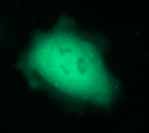 Expressiom of Midoriishi-Cyan in HeLa cells
| Midoriishi-Cyan1 spectrum data files (text files) |
|---|
| Midoriishi-Cyan1 excitation ( |
| Midoriishi-Cyan1 emission ( |
| mMidoriishi-Cyan1 excitation ( |
| mMidoriishi-Cyan1 emission ( |
Note: The file is in a tab-delimited text format. It contains values of the wavelength (0.5nm spacing) and brightness (fluorescence intensity peak value normalized to 1). Use a spreadsheet program to create a spectrum that will help you in choosing the appropriate excitation filter, dichroic mirror and fluorescence filter.
References
※CoralHue™ fluorescent proteins were co-developed with the Laboratory for Cell Function and Dynamics, the Advanced Technology Development Center, the Brain Science Institute, RIKEN. MBL possesses the license and deals in the products.

- Fluorescent protein
- Basic fluorescent proteins
- Midoriishi-Cyan [Outline] [Product list]
- Umikinoko-Green [Outline] [Product list]
- Azami-Green [Outline] [Product list]
- Kusabira-Orange [Outline] [Product list]
- dKeima570 [Outline] [Product list]
- Keima-Red [Outline] [Detection of mitophagy with Keima-Red] [Product list]
- Photoconvertible fluorescent proteins
- Dronpa-Green [Outline] [Product list]
- Kaede [Outline] [Product list]
- Kikume Green-Red [Outline] [Product list]
- Organelle targeting vectors [Product list]
- Advanced fluorescent indicators
- Image based Protein-Protein interaction analysis "Fluoppi" [Outline] [Fluoppi Red] [Product list]
- Cell cycle indicator "Fucci" [Outline] [Product list]
- Protein-Protein Interaction Detection System "CoralHue™ Fluo-chase Kit" [Outline] [Product list]
- Anti-CoralHue™ fluorescent protein antibodies
- Anti-CoralHue™ fluorescent protein antibodies [Outline] [Product list]
- Resources
- CoralHue™ vectors full DNA sequence
- Spectrum