


- Large Stokes Shift
- Unique Spectrum
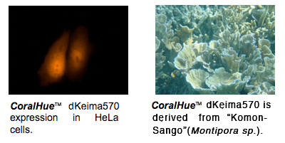
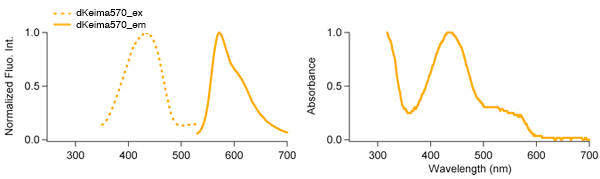
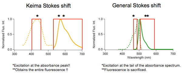
CoralHue™ dKeima570, originally cloned from the stony coral Montipora sp.(Komon-sango in Japanese), absorbs light maximally at 440 nm and emits orange-red light at 570 nm. Because of the extremely large Stokes shift (130 nm) of CoralHue™ dKeima570, the maximum fluorescence can be obtained by the maximum excitation without sacrificing either excitation or fluorescence.
Although several fluorescent proteins have a large Stokes shift, they have green fluorescence as a result of excitation with UV light at around 380 nm. However, the use of such toxic UV light is not suitable for observation in living organisms. Keima is the first red fluorescent protein having a large Stokes shift.
The combination of orange-red emission, a large Stokes shift, stability at 37°C in eukaryotic cells, and being dimeric make CoralHue™ dKeima570 a superb reporter protein for labeling subcellular structures in multicolor fluorescence analyses. The orange-red fluorescence is stable under normal aerobic conditions.
| Keima-Red spectrum data files (text files) | |
|---|---|
| dKeima570 excitation ( |
dKeima570 emission ( |
Note: The file is in a tab-delimited text format. It contains values of the wavelength (1nm spacing) and brightness (fluorescence intensity peak value normalized to 1). Use a spreadsheet program to create a spectrum that will help you in choosing the appropriate excitation filter, dichroic mirror and fluorescence filter.
| CHARACTERISTIC | dKeima570 |
|---|---|
| Oligomerization | Dimer |
| Number of amino acid | 222 |
| Excit./Emiss. maxima (nm) | 440/570 |
| Molar extinction coefficient (M-1cm-1) | 14,000 (492 nm) |
| Fluorescence quantum yield | 0.15 |
| Brightness*1 | 2.1 |
| pH sensitivity | pKa=6.5 |
| Cytotoxicity*2 | No |
*1Brightness: Molar Extinction Coefficient ×Fluorescence Quantum Yield / 1000
*2Toxicity when expressed in HeLa cells
References
- Kogure T, Karasawa S, Araki T, Saito K, Kinjo M, Miyawaki A. A fluorescent variant of a protein from the stony coral Montipora facilitates dual-color single-laser fluorescence cross-correlation spectroscopy. Nat Biotechnol. 2006 (5):577-81. PMID: 16648840
※CoralHue™ fluorescent proteins were co-developed with the Laboratory for Cell Function and Dynamics, the Advanced Technology Development Center, the Brain Science Institute, RIKEN. MBL possesses the license and deals in the products.
Please send us a completed and signed License Acknowledgement.
Please note that the product will be shipped after we receive a
completed and signed License Acknowledgement (PDF file on the right).
Prior to shipping, our staff may contact you to verify the intended use
of the product. Please also note that a separate agreement or a contract
may be required depending on the intended use.
The product you have ordered is sold for research purpose only. It is
not intended for industrial or clinical use, and shall not be used for
any purposes other than research. You are also asked not to hand over or
resell the product to a third party.
If you belong to a profit organization or a business corporation, or
wish to use the product for profit or commercial purposes, please
contact us.

- Fluorescent protein
- Basic fluorescent proteins
- Midoriishi-Cyan [Outline] [Product list]
- Umikinoko-Green [Outline] [Product list]
- Azami-Green [Outline] [Product list]
- Kusabira-Orange [Outline] [Product list]
- dKeima570 [Outline] [Product list]
- Keima-Red [Outline] [Detection of mitophagy with Keima-Red] [Product list]
- Photoconvertible fluorescent proteins
- Dronpa-Green [Outline] [Product list]
- Kaede [Outline] [Product list]
- Kikume Green-Red [Outline] [Product list]
- Organelle targeting vectors [Product list]
- Advanced fluorescent indicators
- Image based Protein-Protein interaction analysis "Fluoppi" [Outline] [Fluoppi Red] [Product list]
- Cell cycle indicator "Fucci" [Outline] [Product list]
- Protein-Protein Interaction Detection System "CoralHue™ Fluo-chase Kit" [Outline] [Product list]
- Anti-CoralHue™ fluorescent protein antibodies
- Anti-CoralHue™ fluorescent protein antibodies [Outline] [Product list]
- Resources
- CoralHue™ vectors full DNA sequence
- Spectrum