


Keima-Red
- Large Stokes shift
- For use with simultaneous multicolor imaging
- For use with single laser line FCCS
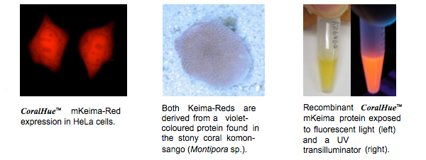
CoralHue™ dimeric Keima-Red (dKeima-Red) and CoralHue™ monomeric
Keima-Red (mKeima-Red) are red fluorescent proteins with extremely large
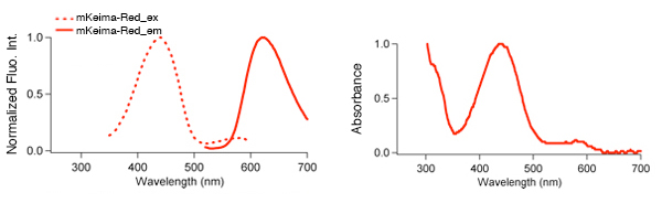
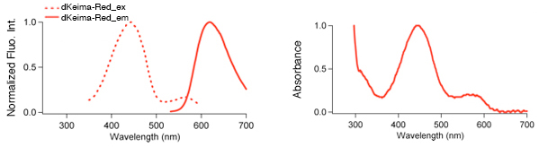
Stokes shift. They absorb light maximally at 440 nm and emit red light at 616
nm and 620 nm, respect ively. There are no other fluorescent proteins with this
unique fluorescence. Because of this characteristic, they are excited by a very
short wavelength but emit a long wavelength. Keima is named after a shogi
(Japanese chess) piece Keima (![]() ) that can move in the hopping manner, similar to the
knight in the game of chess.
) that can move in the hopping manner, similar to the
knight in the game of chess.
The large Stokes shift property of Keima-Red allows effective applications such as for single wavelength excitation simultaneous multi-color imaging and single laser line FCCS.
mKeima-Red
dKeima-Red
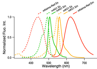
| Keima-Red spectrum data files (text files) | |
|---|---|
| mKeima-Red excitation & emission ( |
dKeima-Red excitation & emission ( |
Note: The file is in a tab-delimited text format. It contains values of the wavelength (1nm spacing) and brightness (fluorescence intensity peak value normalized to 1). Use a spreadsheet program to create a spectrum that will help you in choosing the appropriate excitation filter, dichroic mirror and fluorescence filter.
| CHARACTERISTIC | dKeima-Red | mKeima-Red |
|---|---|---|
| Oligomerization | Dimer | Monomer |
| Number of amino acid | 222 | 222 |
| Excit./Emiss. maxima (nm) | 440/616 | 440/620 |
| Molar extinction coefficient (M-1cm-1) | 24,600 (440 nm) | 14,400 (440 nm) |
| Fluorescence quantum yield | 0.31 | 0.24 |
| Brightness*1 | 7.6 | 3.5 |
| pH sensitivity | pKa=6.5 | pKa=6.5 |
| Cytotoxicity*2 | No | No |
*1Brightness: Molar Extinction Coefficient ×Fluorescence Quantum Yield / 1000
*2Toxicity when expressed in HeLa cells
dKeima-Red and mKeima-Red are useful for labeling organelle.
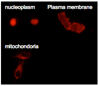 dKeima-Red and mKeima-Red are easily expressed in a wide range of organisms. dKeima-Red and mKeima-Red has been demonstrated in the nucleoplasm, plasma membrane, and mitochondria targeting signal models.
mKeima-Red use in multicolor labeling of mammalian cells.
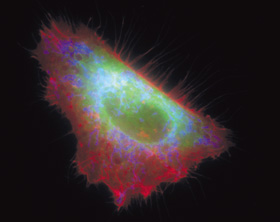 An
image of the HeLa cell with mKO1 localized on the plasma membrane
(red), mAG1 in the endoplasmic reticulum (green) and mKeima-Red in the
mitochondria (blue).
An
image of the HeLa cell with mKO1 localized on the plasma membrane
(red), mAG1 in the endoplasmic reticulum (green) and mKeima-Red in the
mitochondria (blue).
[Fluorescent proteins and their target organella]
mKeima-Red:Mitochondria (blue)
mKO1 (CoralHue™ monomeric Kusabira-Orange):plasma membrane (red)
mAG1 (CoralHue™ monomeric Azami-Green) :endoplasmic reticulum (green)
The image and information are provided by Dr. Miyawaki
Atsushi, Laboratory for Cell Function and Dynamics,
BSI, RIKEN
Filters and mirrors
 mKeima-Red Ex : 440AF21, Em : 610ALP, DM : 590DRLP
mAG1 Ex : BP460-480HQ, Em : BA495-540HQ, DM : DM485
mKO1 Ex : BP520-540HQ, Em : BA555-600HQ, DM : DM545HQ
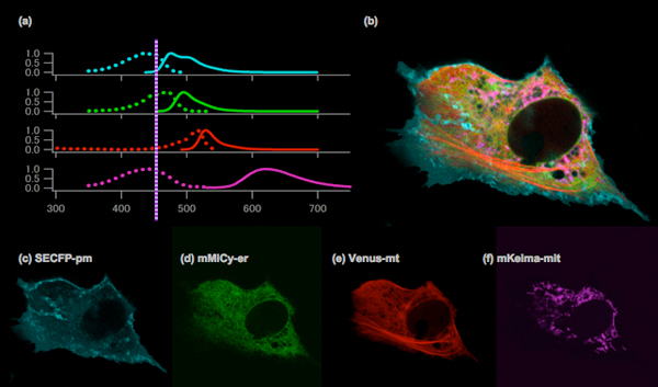
Simultaneous four-color imaging of subcellular structures in a Vero cell using a single laser line (458 nm).
Normalized excitation and emission spectra of SECFP, mMiCy, Venus and mKeima (a). An image of the Vero cell with SECFP localized on the plasma membrane (cyan), mMiCy in the endoplasmic reticulum (green), YFP along the microtubules (red) and mKeima in the mitochondria (purple) (b) . The image was created by merging the following images, which were obtained using spectral imaging: SECFP-pm (c), mMiCy-er (d), Venus-mt (e) and mKeima-mit (f).
Spectra imaging with a single laser line at 458 nm (Ar ion laser) was performed using the 32 channels of the LSM 510 META system (Carl Zeiss).
Images were kindly provided by Takako Kogure, Atsushi Miyawaki (Laboratory for Cell Function and Dynamics , BSI, RIKEN).
Recommended antibodies
CoralHue™ dKeima-Red and mKeima-Red can be recognized using antibodies as shown below.
- Anti-monomeric Keima-Red for WB, Code No. M126-3M
WB: Western blotting, IP: Immunoprecipitation
References
- Katayama H, et al., A sensitive and quantitative technique for detecting autophagic events based on lysosomal delivery. Chem. Biol. 18, 1042-1052 (2011) PMID: 21867919
- Kogure T, et al., A fluorescent variant of a protein from the stony coral Montipora facilitates dual-color single-laser fluorescence cross-correlation spectroscopy. Nat Biotechnol.24: 577-581 (2006) PMID: 16648840
- Kawano H, et al., Two-photon dual-color imaging using fluorescent proteins. Nat Methods. 5(5):373-4. (2008) PMID: 18446153
- Kogure T, et al., Fluorescence imaging using a fluorescent protein with a large Stokes shift. Methods. 45: 223-226 (2008) PMID: 18586106
- Matsumoto Y, et al., Intranuclear fluorescence resonance energy transfer analysis of plasmid DNA decondensation from nonviral gene carriers. J Gene Med. 11: 615-623 (2009) PMID: 19396931
- Violot S, et al., Reverse pH-dependence of chromophore protonation explains the large Stokes shift of the red fluorescent protein mKeima. J Am Chem Soc. 131: 10356-10357 (2009) PMID: 19722611
- Henderson JN, et al., Excited state proton transfer in the red fluorescent protein mKeima. J Am Chem Soc. 131: 13212-13213 (2009) PMID: 19708654
※CoralHue™ fluorescent proteins were co-developed with the Laboratory for Cell Function and Dynamics, the Advanced Technology Development Center, the Brain Science Institute, RIKEN. MBL possesses the license and deals in the products.
Please send us a completed and signed License Acknowledgement.
Please note that the product will be shipped after we receive a
completed and signed License Acknowledgement (PDF file on the right).
Prior to shipping, our staff may contact you to verify the intended use
of the product. Please also note that a separate agreement or a contract
may be required depending on the intended use.
The product you have ordered is sold for research purpose only. It is
not intended for industrial or clinical use, and shall not be used for
any purposes other than research. You are also asked not to hand over or
resell the product to a third party.
If you belong to a profit organization or a business corporation, or
wish to use the product for profit or commercial purposes, please
contact us.

- Fluorescent protein
- Basic fluorescent proteins
- Midoriishi-Cyan [Outline] [Product list]
- Umikinoko-Green [Outline] [Product list]
- Azami-Green [Outline] [Product list]
- Kusabira-Orange [Outline] [Product list]
- dKeima570 [Outline] [Product list]
- Keima-Red [Outline] [Detection of mitophagy with Keima-Red] [Product list]
- Photoconvertible fluorescent proteins
- Dronpa-Green [Outline] [Product list]
- Kaede [Outline] [Product list]
- Kikume Green-Red [Outline] [Product list]
- Organelle targeting vectors [Product list]
- Advanced fluorescent indicators
- Image based Protein-Protein interaction analysis "Fluoppi" [Outline] [Fluoppi Red] [Product list]
- Cell cycle indicator "Fucci" [Outline] [Product list]
- Protein-Protein Interaction Detection System "CoralHue™ Fluo-chase Kit" [Outline] [Product list]
- Anti-CoralHue™ fluorescent protein antibodies
- Anti-CoralHue™ fluorescent protein antibodies [Outline] [Product list]
- Resources
- CoralHue™ vectors full DNA sequence
- Spectrum