


Dronpa-Green
- Photoswitchable fluorescent protein
- Reversible "on-off" switching
- Useful for molecular dynamics investigation
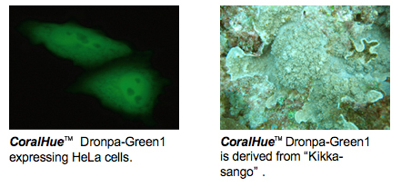
The fluorescent protein Dronpa gene was originally cloned from stony coral Echinophyllia sp. (Kikka-sango in Japanese).
CoralHue™ Dronpa-Green1 (DG1) is a monomeric green fluorescent protein that is
photochromic: its fluorescence is reversible, with "on-off" switching by demand
through exposure to di fferent wavelengths of light. DG 1 bleaches immediately
following strong excitation at -500 nm. Bleached DG1 completely regains its bright
green fluorescence after irradiation at -400 nm. This unique property of DG1 is useful
for repeated measurements of mobility dynamics (e.g. diffusion, transport) of
fluorescent-labeled molecules in living cells.
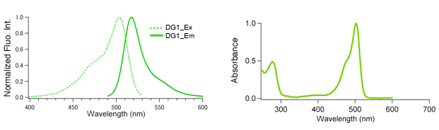
DG1
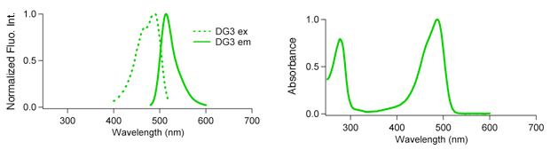
DG3
| Dronpa-Green spectrum data files (text files) | |
|---|---|
| Dronpa-Green excitation ( |
Dronpa-Green emission ( |
| Dronpa-Green3 excitation ( |
Dronpa-Green3 emission ( |
Note: The file is in a tab-delimited text format. It contains values of the wavelength (0.5nm spacing) and brightness (fluorescence intensity peak value normalized to 1). Use a spreadsheet program to create a spectrum that will help you in choosing the appropriate excitation filter, dichroic mirror and fluorescence filter.
| CHARACTERISTIC | Dronpa-Green1 | Dronpa-Green3 |
|---|---|---|
| Oligomerization | Monomer | Monomer |
| Number of amino acid | 225 | 225 |
| Excit./Emiss. maxima (nm) | 503/518 | 491/514 |
| Molar extinction coefficient (M-1cm-1) | 95,000 (503 nm) | 58,000 (487 nm) |
| Fluorescence quantum yield | 0.85 | 0.28 |
| Brightness*1 | 80.7 | 16.2 |
| pH sensitivity | pKa=5.0 | pKa=5.0 |
| Cytotoxicity*2 | No | No |
*1Brightness: Molar Extinction Coefficient ×Fluorescence Quantum Yield / 1000
*2Toxicity when expressed in HeLa cells
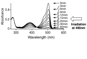
Spectra change of Dronpa
The absorption spectrum of Dronpa at pH7.4 displays a major peak at 503 nm and a minor peak at 390 nm. The absorbance of Dronpa at pH 7.4 were measured during continuous illumination at 490 nm ± 10. After a 40-min incubation, nearly all the Dronpa molecules had been converted into a neutral, nonfluorescent state.
Ando, R., Mizuno, H. and Miyawaki, A.(2004) Science 306, 1370-1373.
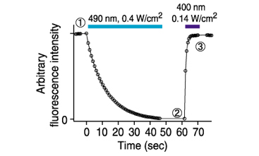
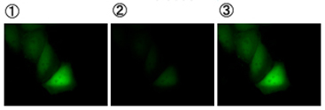
Photochoromic properties of Dronpa
Dronpa was transfected into HeLa cells followed by fixation and the time course of fluorescence was monitored. Photoactivation of DG1 required much less photon energy than did photobleaching, with respective quantum yields of 0.37 (ΦPA) and 0.00032 (ΦPB).
Ando, R., Mizuno, H. and Miyawaki, A.(2004) Science 306, 1370-1373.
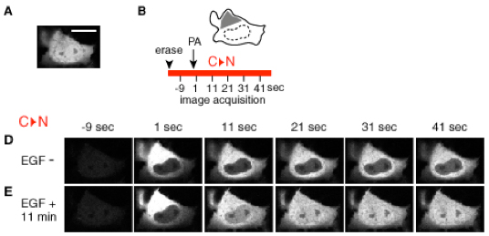
Monitoring the nuclear import of ERK1-Dronpa in Cos7 cells
(A) Confocal image of a COS7 cell expressing ERK1-Dronpa. (B) Schematic of the cell shown in A. The regions illuminated with the 405-nm laser line are shown in gray. The illuminated regions were inside the cytosol (left) or nucleus (right) to monitor nuclear import (C→N). The time periods monitored for nuclear import are shown in red. (D and E) Nuclear import of ERK1-Dronpa before stimulation of D or after an 11-min incubation with EGF (E).
Ando, R., Mizuno, H. and Miyawaki, A.(2004) Science 306, 1370-1373.
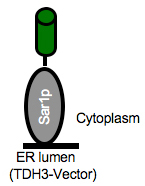
Molecular imaging of Sar1P-Dronpa in yeast
CoralHue™ Dronpa-Green1 is useful for monitoring molecular dynamics. Sar1P, of a small G protein family, was fused with Dronpa-Green1 (Sar1P-Dronpa). Sar1P-Dronpa was expressed in yeast. The videos show Sar1P-Dronpa monitoring molecular dynamics in the mother cell and daughter cells.
Provided by Dr. Kurokawa and Dr. Nakano
Live Cell Molecular Imaging Research Team, ASI, RIKEN
Mother cell → Daughter cell
|
daughter cell → Mother cell
|
|
|
|
|
 |
References
- Ando R, Mizuno H, Miyawaki A. Regulated fast nucleocytoplasmic shuttling observed by reversible protein highlighting. Science. (2004) 306: 1370-3. PMID: 15550670
- Ando R, Flors C, Mizuno H, Hofkens J, Miyawaki A. Highlighted generation of fluorescence signals using simultaneous two-color irradiation on Dronpa mutants. Biophys J. (2007) 92:L97-9. PMID: 17384059
- Habuchi S, Ando R, Dedecker P, Verheijen W, Mizuno H, Miyawaki A, Hofkens J. Reversible single-molecule photoswitching in the GFP-like fluorescent protein Dronpa. Proc Natl Acad Sci U S A. (2005) 102:9511-6. PMID: 15972810
- Habuchi S, Dedecker P, Hotta J, Flors C, Ando R, Mizuno H, Miyawaki A, Hofkens J. Photo-induced protonation/deprotonation in the GFP-like fluorescent protein Dronpa: mechanism responsible for the reversible photoswitchingPhotochem Photobiol Sci. (2006) 5:567-76. PMID: 16761085
※CoralHue™ fluorescent proteins were co-developed with the Laboratory for Cell Function and Dynamics, the Advanced Technology Development Center, the Brain Science Institute, RIKEN. MBL possesses the license and deals in the products.
Please send us a completed and signed License Acknowledgement.
Please note that the product will be shipped after we receive a
completed and signed License Acknowledgement (PDF file on the right).
Prior to shipping, our staff may contact you to verify the intended use
of the product. Please also note that a separate agreement or a contract
may be required depending on the intended use.
The product you have ordered is sold for research purpose only. It is
not intended for industrial or clinical use, and shall not be used for
any purposes other than research. You are also asked not to hand over or
resell the product to a third party.
If you belong to a profit organization or a business corporation, or
wish to use the product for profit or commercial purposes, please
contact us.

- Fluorescent protein
- Basic fluorescent proteins
- Midoriishi-Cyan [Outline] [Product list]
- Umikinoko-Green [Outline] [Product list]
- Azami-Green [Outline] [Product list]
- Kusabira-Orange [Outline] [Product list]
- dKeima570 [Outline] [Product list]
- Keima-Red [Outline] [Detection of mitophagy with Keima-Red] [Product list]
- Photoconvertible fluorescent proteins
- Dronpa-Green [Outline] [Product list]
- Kaede [Outline] [Product list]
- Kikume Green-Red [Outline] [Product list]
- Organelle targeting vectors [Product list]
- Advanced fluorescent indicators
- Image based Protein-Protein interaction analysis "Fluoppi" [Outline] [Fluoppi Red] [Product list]
- Cell cycle indicator "Fucci" [Outline] [Product list]
- Protein-Protein Interaction Detection System "CoralHue™ Fluo-chase Kit" [Outline] [Product list]
- Anti-CoralHue™ fluorescent protein antibodies
- Anti-CoralHue™ fluorescent protein antibodies [Outline] [Product list]
- Resources
- CoralHue™ vectors full DNA sequence
- Spectrum