
- Japan(Japanese / English)
- Global
- MBL TOP
- MBL site search


Azami-Green
- Bright green fluorescence
- High pH stability
- Monomer/tetramer
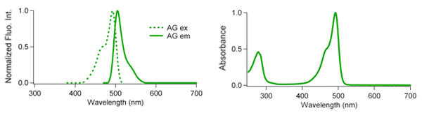
CoralHue™ Azami-Green (AG), cloned from the stony coral Galaxea fascicularis (Azami-sango in Japanese), absorbs light maximally at 492 nm and emits green light at 505 nm. AG is stable in the biological pH range and does not show a significant loss of fluorescent signal, making it advantageous over other fluorescent proteins such as EGFP. And AG also matures rapidly to form tetramers that are brighter than EGFP.
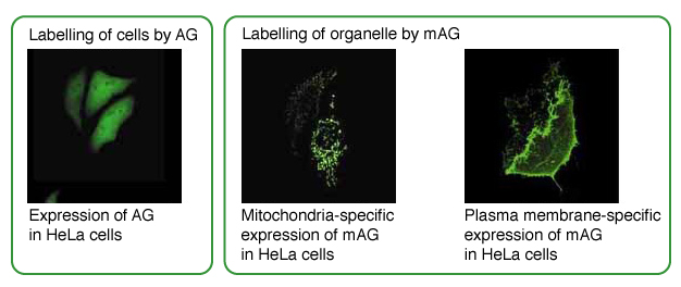
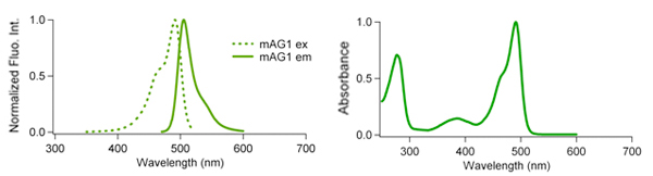
CoralHue™ monomeric Azami-Green (mAG1) maintains the brightness and pH stability of the parent protein AG. mAG1 can be used for labeling proteins or subcellular structures.
AG
mAG
| Azami Green(AG) spectrum data files (text files) | |
|---|---|
| AG excitation ( |
AG emission ( |
| mAG1 excitation ( |
mAG1 emission ( |
Note: The file is in a tab-delimited text format. It contains values of the wavelength (0.5nm spacing) and brightness (fluorescence intensity peak value normalized to 1). Use a spreadsheet program to create a spectrum that will help you in choosing the appropriate excitation filter, dichroic mirror and fluorescence filter.
| CHARACTERISTIC | AG | mAG1 |
|---|---|---|
| Oligomerization | Tetramer | Monomer |
| Number of amino acid | 225 | 225 |
| Excit./Emiss. Maxima (nm) | 492/505 | 492/505 |
| Molar extinction coefficient (M-1cm-1) | 72,300 (492 nm) | 55,500 (492 nm) |
| Fluorescence quantum yield | 0.67 | 0.74 |
| Brightness *1 | 48.4 | 41.0 |
| pH sensitivity | pKa<5.0 | pKa=5.8 |
| Cytotoxicity *2 | No | No |
*1Brightness: Molar Extinction Coefficient ×Fluorescence Quantum Yield / 1000
*2Toxicity when expressed in HeLa cells
AG is resistant to acidity
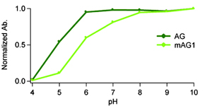 AG shows high resistance in a wide range of biological pH. This allows AG to be used identify cells or to report gene expression without being influenced by pH change. The pH dependence of both CoralHue™ AG and mAG1 was identical to that of its absorption spectra.
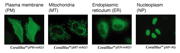
AG and mAG1 are useful for labeling organelle.
AG and mAG1 performance in fusions has been demonstrated in the nucleoplasm, plasma membrane, ER and mitochondria-targeting signal models. We offer these constructs as targeting vector series.
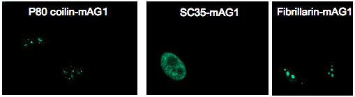
mAG1 is available for protein labeling
mAG1 performance in fusion has been demonstrated in p80 coilin, SC35 and fibrillarin.
Recommended antibodies
CoralHue™ AG and mAG1 can be recognized using antibodies as shown below.
WB: Western blotting, IP: Immunoprecipitation, IC: Immunocytochemistry, IH: Immunohictochemistry
References
- Karasawa S, Araki T, Yamamoto-Hino M, Miyawaki A, A green-emitting fluorescent protein from Galaxeidae coral and its monomeric version for use in fluorescent labeling; J Biol Chem. 278, 34167-34171 (2003) PMID: 12819206
- Ebisawa T, Yamamura A, Kameda Y, Hayakawa K, Nagata K, Tanokura M. Crystallization and preliminary X-ray analysis of a monomeric
mutant of Azami-Green (mAG), an Aequorea victoria green fluorescent
protein-like green-emitting fluorescent protein from the stony coral
Galaxea fascicularis. Acta Crystallogr Sect F Struct Biol Cryst Commun. 65(Pt 12):1292-5. (2009) PMID: 20054132
※CoralHue™ fluorescent proteins were co-developed with the Laboratory for Cell Function and Dynamics, the Advanced Technology Development Center, the Brain Science Institute, RIKEN. MBL possesses the license and deals in the products.
Please send us a completed and signed License Acknowledgement.
Please note that the product will be shipped after we receive a
completed and signed License Acknowledgement (PDF file on the right).
Prior to shipping, our staff may contact you to verify the intended use
of the product. Please also note that a separate agreement or a contract
may be required depending on the intended use.
The product you have ordered is sold for research purpose only. It is
not intended for industrial or clinical use, and shall not be used for
any purposes other than research. You are also asked not to hand over or
resell the product to a third party.
If you belong to a profit organization or a business corporation, or
wish to use the product for profit or commercial purposes, please
contact us.
Pick Up products

- Fluorescent protein
- Basic fluorescent proteins
- Midoriishi-Cyan [Outline] [Product list]
- Umikinoko-Green [Outline] [Product list]
- Azami-Green [Outline] [Product list]
- Kusabira-Orange [Outline] [Product list]
- dKeima570 [Outline] [Product list]
- Keima-Red [Outline] [Detection of mitophagy with Keima-Red] [Product list]
- Photoconvertible fluorescent proteins
- Dronpa-Green [Outline] [Product list]
- Kaede [Outline] [Product list]
- Kikume Green-Red [Outline] [Product list]
- Organelle targeting vectors [Product list]
- Advanced fluorescent indicators
- Image based Protein-Protein interaction analysis "Fluoppi" [Outline] [Fluoppi Red] [Product list]
- Cell cycle indicator "Fucci" [Outline] [Product list]
- Protein-Protein Interaction Detection System "CoralHue™ Fluo-chase Kit" [Outline] [Product list]
- Anti-CoralHue™ fluorescent protein antibodies
- Anti-CoralHue™ fluorescent protein antibodies [Outline] [Product list]
- Resources
- CoralHue™ vectors full DNA sequence
- Spectrum