
- Japan(Japanese / English)
- Global
- MBL TOP
- MBL site search


Labels for detecting antibodies
Antibodies are labeled with various substances for detection.
When using a commercial kit to label antibodies, care must be taken not to lose the activity of the antibody.
Common types of labels
・Fluoresecent labeling
・Enzyme labeling
・Biotin
・Magnetic beads, agarose beads, magnetic agarose beads
・Colloidal gold
Fluoresecent labeling
Fluorescent labeled antibodies are used for flow cytometry and immunofluorescence staining. Multiple antibodies with different fluorescent labels can be used for double or triple staining.
| Type of fluorophore | |
|---|---|
| Fluorescent dye (low molecular weight fluorescent dye) | FITC (fluorescein isothiocyanate), Alexa Fluor® dye Cy dyes, etc. |
| Fluorescent protein | PE (phycoerythrin), APC (allophycocyanin), etc. |
Application example of fluorescent-labeled antibody: Immunofluorescence staining
Immunofluorescence staining with an antibody to an organelle marker
HeLa cells were stained with an antibody to the Golgi protein GM130.
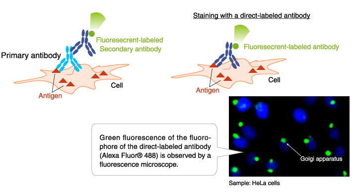
Antibody:Anti-GM130 mAb-Alexa Fluor® 488(Code No. M179-A48)
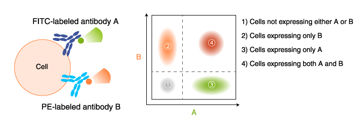
Application example of fluorescent-labeled antibody: Flow cytometry
T cells are separated by type using fluorescent-labeled antibodies to cell surface markers.
T cells are separated into helper T cells, cytotoxic T cells, etc., by flow cytometry using antibodies labeled with different fluorophores (CD4-FITC, CD8-PE).
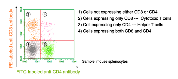
PE-labeled anti-CD8 antibody (Clone: KT15, Code No. D271-5)
FITC-labeled anti-CD4 antibody (Clone: GK1.5, Code No. D341-4)
Enzyme labeling
Enzyme-labeled antibodies are used for ELISA, Western blotting, and immunostaining.
Labeled antibodies are detected by reaction with a substrate that emits light or changes color.
| Enzyme name | Chromogenic substrate | Chemiluminescence substrate |
|---|---|---|
| HRP(Horseradish peroxidase) | DAB and TMB | Luminol-based (ECL) |
| AP(Alkaline phosphatase) | BCIP/NBT and pPNPP | Dioxetane-based (CDP-star®) |
Application example of enzyme-labeled antibodies: Immunohistochemical staining
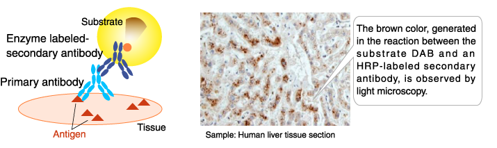
The result of immunohistochemical staining indicates that autophagy was induced in the liver tissue.
Application example of enzyme-labeled antibodies: Western blotting
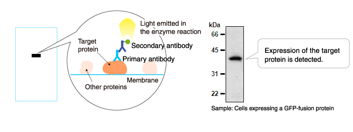
Primary antibody: Mouse anti-GFP antibody (Clone: 1E4, Code No. M048-3)
Secondary antibody: Anti-mouse IgG antibody (HRP-labeled, Code No. 330)
Biotin
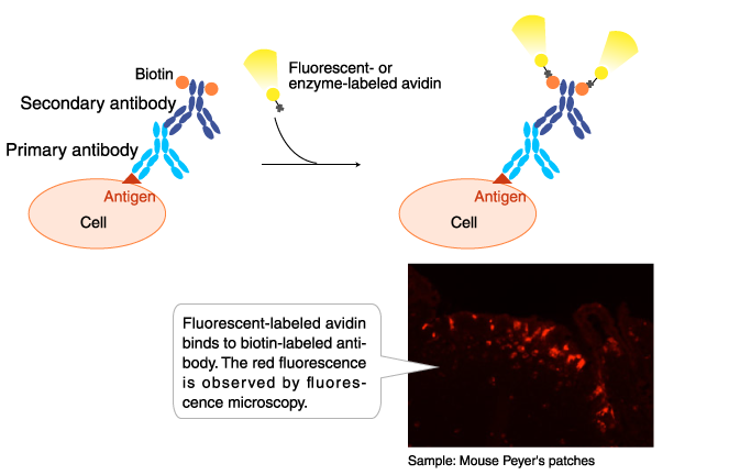
Biotin-labeled antibodies are used in flow cytometry, immunofluorescence staining, and immunohistochemical staining. Biotin-labeled antibodies are detected by reaction with dye-, enzyme-, or fluorophore-labeled avidin.
Antibody: Biotin-labeled anti-GP2 (Glycoprotein 2) antibody (Code No. D278-6)
Agarose, magnetic beads, magnetic agarose beads
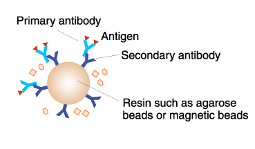
Antibody-conjugated resins, such as magnetic beads and agarose, are used for immunoprecipitation.
>>The principle and method of immunoprecipitation (IP)
Colloidal gold
Gold-labeled antibodies are used in immunoelectron microscopy. This method is highly sensitive, and the signal does not fade as in fluorescent-labeling and enzyme-labeling.
Immunoelectron micrograph of autophagosomes detected using the marker LC3
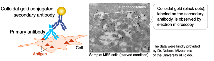
Primary antibody: Anti-LC3 monoclonal antibody (Clone: 4E12, Code No. M152-3)
| Fluorescent | FITC-labeled antibodies PE-labeled antibodies Alexa Fluor® 488-labeled antibodies Alexa Fluor® 594-labeled antibodies Alexa Fluor® 647-labeled antibodies |
| Enzyme | HRP-labeled antibodies AP-labeled antibodies |
| Other |
Biotin-labeled antibodies Antibody-conjugated magnetic beads Antibody-conjugated magnetic agarose Primary antibody-conjugated agarose |